Optical in vivo imaging technology mainly uses bioluminescence and fluorescence. Bioluminescence is the labeling of cells or DNA with the luciferase gene, while fluorescent techniques are labeled with fluorescent reporter groups (GFP, RFP, Cyt and dyes, etc.). Visible in vivo imaging records the movement and changes of the same observation target (labeled cells and genes) by recording the same group of subjects at different time points, and the obtained data is more authentic. Due to its extremely simple operation, intuitive results, and high sensitivity, it has been widely used in life science, medical research and drug development in recent years.
Researchers such as Hye-Min Kang from Kyung Hee University in Korea used VISQUE in vivo imaging technology to study the changes of blood vessel structure and blood flow in mice at different ages.
First, the researchers used the indocyanine green (ICG) fluorescent tracer to give mice a tail vein injection, using real-time imaging mode, exposure time 160ms, interval 2min, a total of 50 real-time images were taken, by selecting the right hemisphere The routine indicators of regional dynamics, Trixing, Imax, BFI, and MTT, found that the blood flow intensity of the heads of mice at different ages did differ (Fig. 1), indicating that the young mice had faster venous blood flow and the brain. Blood volume is also greater.
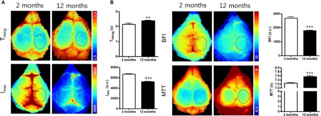
Figure 1: Comparison of cerebral blood flow between 2 month old mice and 12 months old mice
Next, the researchers continued to observe the distribution of meningeal arteries in the two groups of mice in ICG mode. It was found that the soft cerebrovascular artery of young mice was straight and sharp, and obvious small branches were observed; however, the meningeal arteries of old mice were blunt and tortuous, and the small branches were unclear. At the same time, a single pixel of the same position of the middle and superior sagittal sinus of the two groups of mice was taken, and the corresponding blood flow curve was observed. It was also found that young mice were more advantageous in the detection of the index (Figure 2, Figure 3). These results indicate that young mice have a wider distribution of meningeal arteries and a greater blood volume than older mice.
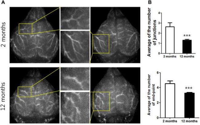
Figure 2: Comparison of the morphology of soft-membrane arteries between 2-month-old mice and 12-month-old mice
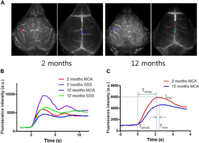
Figure 3: Comparison of cerebral artery (MCA) and superior sagittal sinus (SSS) kinetics in 2 month old mice and 12 month old mice
Finally, the author of the article calculates the blood flow characteristics (Trising, IMAX, BFI, MTT) in the same ROI region of the whole brain through brain blood flow imaging. It is concluded that the blood flow and blood of the young mice are higher than those of the old mice. Flow velocity, blood volume, etc. all have obvious advantages (Figure 4).
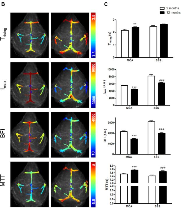
Figure 4: Schematic diagram of blood flow in the middle cerebral artery (MCA) and superior sagittal sinus (SSS) of 2 month old mice and 12 month old mice
VISQUE in vivo imaging technology is playing an increasingly important role in the fields of blood system, tumor, stem cell and pharmacokinetics with its ultra-high resolution and superior pharmacokinetic analysis. It has also become a basic research in biomedicine. An indispensable tool for scientific research.
Document name:
Kang, HM, et al., Age-related changes in pial arterial structure and blood flow in mice. Neurobiol Aging, 2016. 37 : p. 161-170.
Painlessly Silicone-Faced Dressing,Silicone-Faced Dressing,Comfortable Silicone-Faced Dressing,Flexible Silicone-Faced Dressing
Zhende Medical Co.,Ltd , https://www.zhendemedicals.com