Release date: 2016-06-17
Whether it is to replace experimental animals in preclinical studies of drug development or for organ transplantation, artificial organs and tissues have very attractive application prospects. However, artificial organs and tissues constructed in vitro need to survive and perform biological functions in vitro and in vivo. In addition to considering the immune rejection of the body, another problem cannot be ignored. The supply of oxygen and nutrients from artificial to deep surface of artificial organs is gradually decreasing. The trend is that necrosis may occur in cells and tissues at its center. Therefore, the vital organs such as the liver, heart and so on to achieve in vitro preparation and in vivo transplantation survival, whether to build an effective vascular network system has become one of the main limiting factors, but also a bottleneck hindering its clinical application.
At present, the bio-3D printing method is usually used to reserve or accurately locate the inside of the stent to directly print the micro-vascular network structure with good porosity and height, which realizes a high-precision microstructure to a certain extent and realizes rapidly. Vascularization target. However, due to the nature of the matrix material, biocompatibility and mechanical properties, biological 3D printing is not able to meet the actual needs. 3D stamping technology (3D stamping) can provide a new tissue scaffold for tissue engineering by pre-preparing a patterned single layer and forming a three-dimensional vascularized network structure through precise design and layer-by-layer assembly. And vascularization network construction ideas.

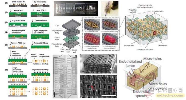
According to a recent report by Nature Materials, the Milica Radisic team at the University of Toronto in Canada developed a built-in vascular network stent chip necessary for human tissue growth using 3D stamping technology and named it AngioChip. It uses a biodegradable, elastic, ultraviolet polymerizable and rapid prototyping POMaC [poly(octamethylene maleate (anhydride) citrate)] polymer material. The patterned POMaC biodegradation layer was prepared by computer software and printed like a seal, and 1D tube, 2D branch catheter and 3D dendritic network-like vascular structure. The patterned POMaC layer is precisely layered and embedded in the microvascular structure, and can be polymerized together by ultraviolet light to form complex microstructures and internal pores. A three-dimensional vascularized tissue scaffold chip was prepared by perfusing the extracellular matrix inside and implanting human active cells. (Biodegradable scaffold with built-in vasculature for organ-on-a-chip engineering and direct surgical anastomosis. Nature Mater., 2016, 15, 669-678, DOI: 10.1038/nmat4570)

The walls of the AngioChip's built-in blood vessels are thin and flexible, and have sufficient mechanical strength to support the perfusion vasculature in the contracted tissue. The vessel wall contains micropores in the micro and nano scales that facilitate molecular exchange and cell migration. The lattice-like matrix scaffold with rich internal connectivity can well simulate the vascular bed, and can grow various active cells in human organs inside. The researchers successfully prepared functional and vascularized liver and heart tissue.
The researchers also implanted this vascularized stent directly into the hind limbs of adult rats for the blood flow of arterial-arterial and arterial-venous vessels in the femoral vessels during surgical anastomosis. The results of the study showed that the blood of the mice flowed smoothly in the AngioChip, and the flow in the natural blood vessels was no different. This attempt gives an effective means of tissue regeneration, which can be directly and quickly translated into in vivo experimental verification.
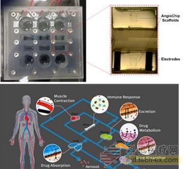
AngioChip can greatly promote the research of organ-on-a-chip. The organ chip is a microfluidic chip biomimetic system based on micromachining technology that simulates the complex microstructure, microenvironment and physiological functions of specific organs of the human body. It is also called microphysiological system or human body (human-on-a). -chip). It is favored by pharmaceutical giants because it is expected to reduce or eventually replace animal models. Since the US President Barack Obama announced the launch of a special research on human body chips jointly established by NIH, FDA and the Ministry of Defense in 2011, the research boom of this new generation of drug evaluation platform technology has been rapidly developed worldwide. The liver and heart chips constructed by AngioChip can also be connected through this embedded artificial vascular access to simulate the biomimetic circulatory system and evaluate its interaction with each other.

AngioChip addresses the vascularization of a key constraint in tissue engineering, organ chips and organ transplantation, providing a good platform for the preparation and culture of three-dimensional vascularized tissue. Researchers are also currently undergoing technology upgrades, from manual assembly to automated production, and developing their commercial application value.
Source: X-MOL
Erythritol For Juice,Organic Erythritol For Juice,Liquid Erythritol,Puresweet Erythritol
Ningxia Eppen Biotec CO.,LTD , https://www.nxeppen.com